Advancing Cancer Diagnostics with Histopathological Cancer Detection on Matrice

Histopathological cancer detection is a pivotal task in medical diagnostics, enabling early identification of cancerous cells in microscopic tissue images. AI-powered computer vision can accurately classify and localize cancerous regions in real-time, supporting pathologists and improving patient outcomes.
This blog details how Matrice enables no-code AI deployment for histopathological cancer detection using deep learning, covering:
Dataset Preparation
Dataset Annotation
Model Training
Model Evaluation
Model Inference
Model Deployment
Dataset Preparation
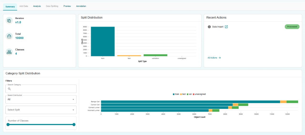
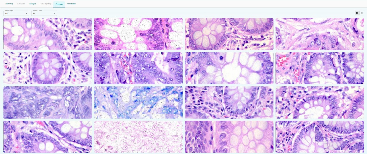
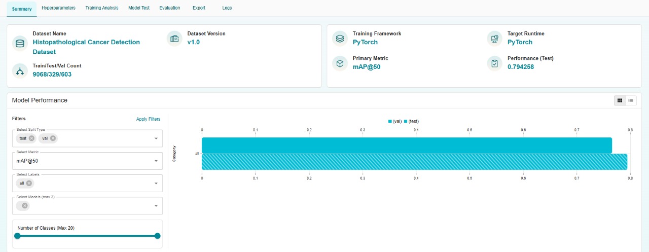
The dataset for this project comprises 10,000 high-resolution microscopic image samples of histopathological slides, annotated to identify cancerous and non-cancerous regions. The dataset is partitioned to ensure robust model development and evaluation:
Total Samples: 10,000
Training Set: 9,068
Validation Set: 603
Testing Set: 329
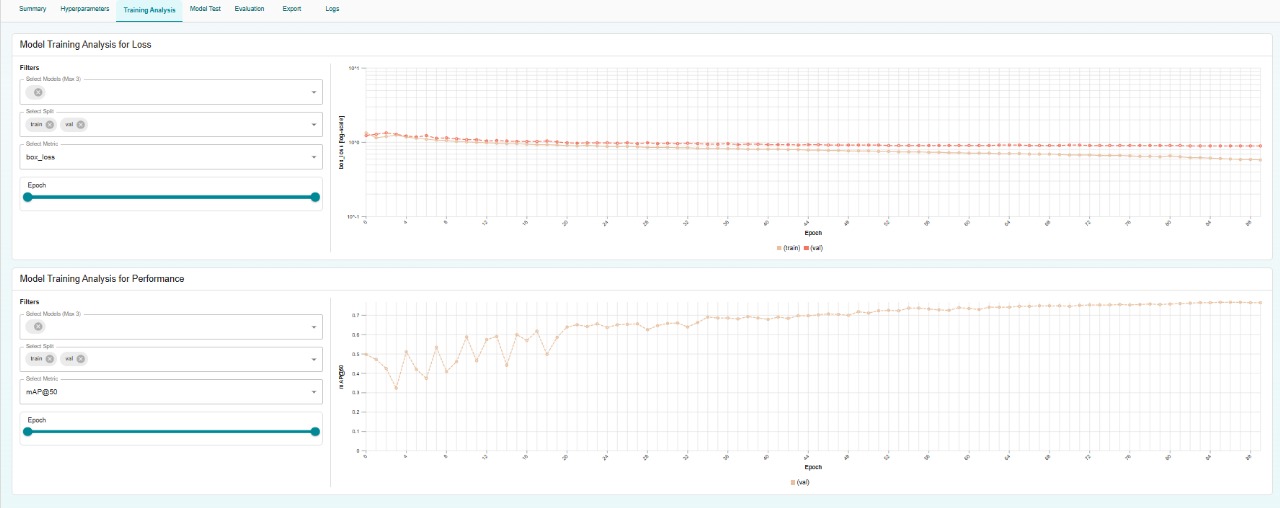
Model Training
We utilized a YOLO-based object detection model to detect and classify cancerous regions in histopathological images. YOLOv8 was selected for its precision and efficiency, ideal for analyzing complex microscopic imagery.
Model: YOLOv8 (multi-class object detection)
Batch Size: 16
Epochs: 90
Learning Rate: 0.001
Optimizer: Auto
Momentum: 0.95
Weight Decay: 0.0005
Model Evaluation
The trained model was evaluated on the test dataset, using key metrics to assess its effectiveness in detecting cancerous regions.
Metric |
Value (Test) |
|---|---|
mAP@50 |
0.79 |
mAP@50-95 |
0.58 |
Recall |
0.77 |
Precision |
0.77 |
Model Inference
The trained model supports export to multiple formats, enabling deployment across various platforms, from hospital diagnostic systems to cloud-based analysis tools.
Supported formats include:
PyTorch (.pt)
ONNX
TensorRT
OpenVINO
This flexibility ensures compatibility with diverse deployment scenarios, such as clinical workstations, telemedicine platforms, or research environments.
Model Deployment
Using Matrice, the trained model can be deployed seamlessly via a no-code interface. Matrice supports:
Real-time inference
API-based integration
Visual dashboards for monitoring
Applications include:
Automated cancer detection in histopathological slides
Decision support for pathologists
Integration into telemedicine and diagnostic platforms
Conclusion
AI-powered histopathological cancer detection enhances diagnostic accuracy and efficiency, supporting early intervention and better patient outcomes. With Matrice, deploying such solutions is streamlined, enabling rapid integration into medical workflows with reduced costs. By leveraging YOLOv8 and a comprehensive dataset, you can develop high-performing models to transform cancer diagnostics with visual intelligence.
Think CV, Think Matrice
Experience 40% faster deployment and slash development costs by 80%