X-Ray Classification for Pneumonia Detection: Speeding Up Diagnosis with AI

Pneumonia can be fatal, and saving lives depends on prompt diagnosis. However, conventional techniques for identifying pneumonia from chest X-rays are frequently laborious and rely on skilled radiologists. AI comes into play. By utilizing machine learning, we can rapidly and precisely identify pneumonia in X-ray pictures, reducing the time needed for diagnosis and guaranteeing patients receive treatment more swiftly. This blog examines how pneumonia detection is being revolutionized and healthcare outcomes are being enhanced by AI-driven X-ray classification.
Comprehending Computer Vision for the Identification of Pneumonia
Computer Vision’s Function
Machines can now examine chest X-rays and detect symptoms of pneumonia, such as fluid accumulation or lung opacity, thanks to computer vision (CV). By automating the detection process, this technology makes it possible to identify patients more quickly and accurately, allowing medical professionals to take prompt action.
Principal Advantages of Pneumonia Detection:
Fast Analysis: The diagnostic procedure can be accelerated by processing X-rays in a matter of seconds.
Early Detection: The technology detects pneumonia symptoms early, allowing for prompt treatment.
Non-Intrusive: By using X-rays to diagnose pneumonia, more intrusive testing is not required.
Consistency: Automated systems minimize human error by producing dependable and repeatable results.
Effect on Health Care Diagnosis
There are various benefits to using automated techniques for pneumonia detection:
Faster Diagnosis: Decisions and treatment can be made more quickly when X-rays are processed quickly.
Increased Accuracy: Automated analysis guarantees more accurate and reliable pneumonia detection.
Enhanced Efficiency: These solutions free up medical personnel to concentrate on complex situations by managing repetitive chores.
Improved Patient Care: Patients receive faster treatments and greater results when diagnoses are made promptly and accurately.
Implementing Pneumonia Detection using Matrice.
This post describes the important stages for building a strong pneumonia detection classification model using Matrice.
Dataset Preparation
Dataset Import
Model Configuration and Training
Model Evaluation
Model Inference
Model Deployment
Step 1: Dataset Preparation
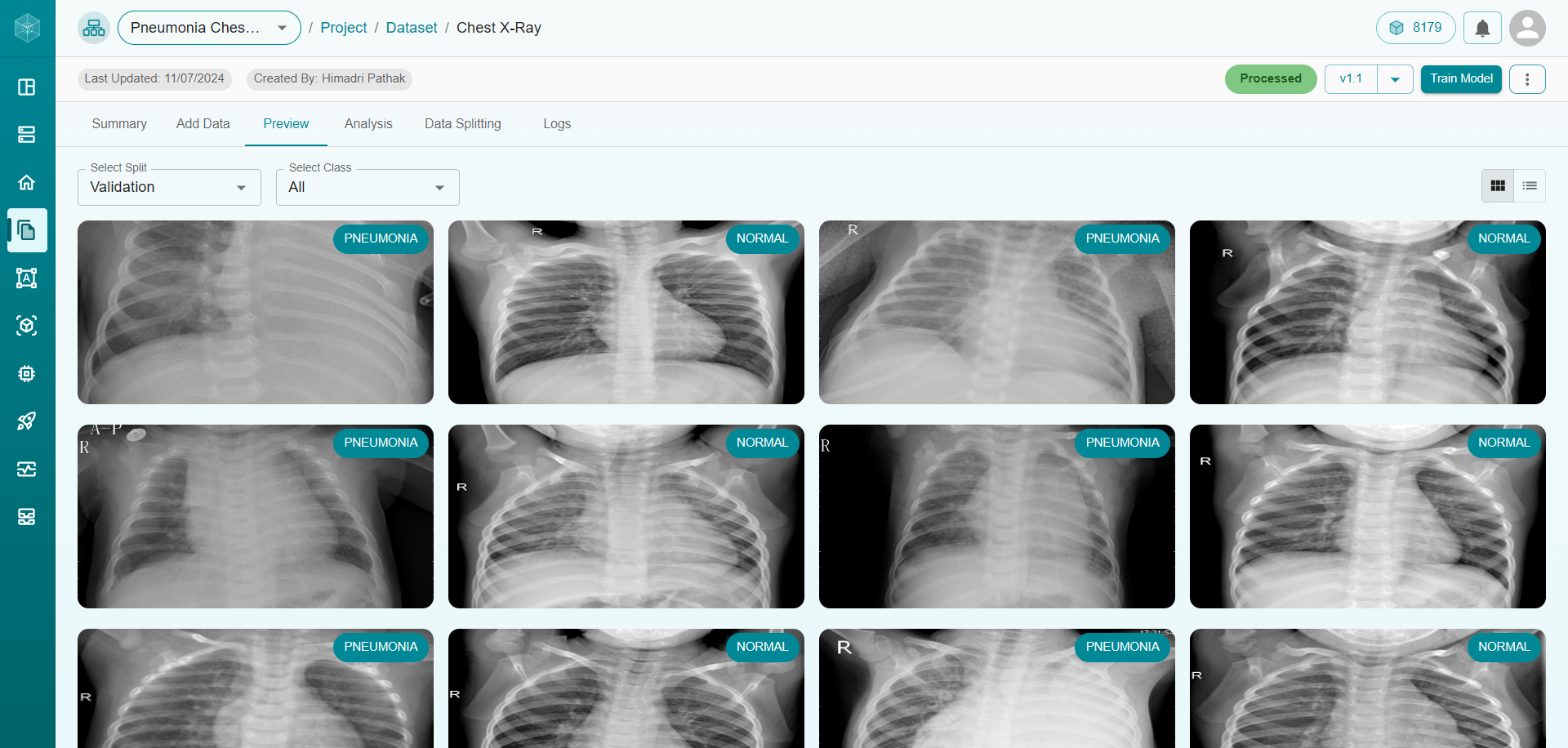
A well-organized dataset is essential for accurate pneumonia detection. Our dataset consisted of tagged chest X-rays that were annotated to identify the picture as Pneumonia or normal. The following steps were used to prepare the dataset:
Data Collection: We obtained chest X-rays from both pneumonia patients and healthy persons who showed no indications of illness.
Data Annotation: Each X-ray was annotated to identify whether it included indications of pneumonia or was a normal, healthy lung.
Data Splitting: The dataset was divided into training (59.99%), testing (25%), and validation (15.01%) subsets to ensure the model’s ability to generalize across diverse cases.
Step 2: Dataset Import
Matrice facilitates the uploading and organization of medical imaging datasets. This stage involved:
Step 3: Model Configuration and Training.
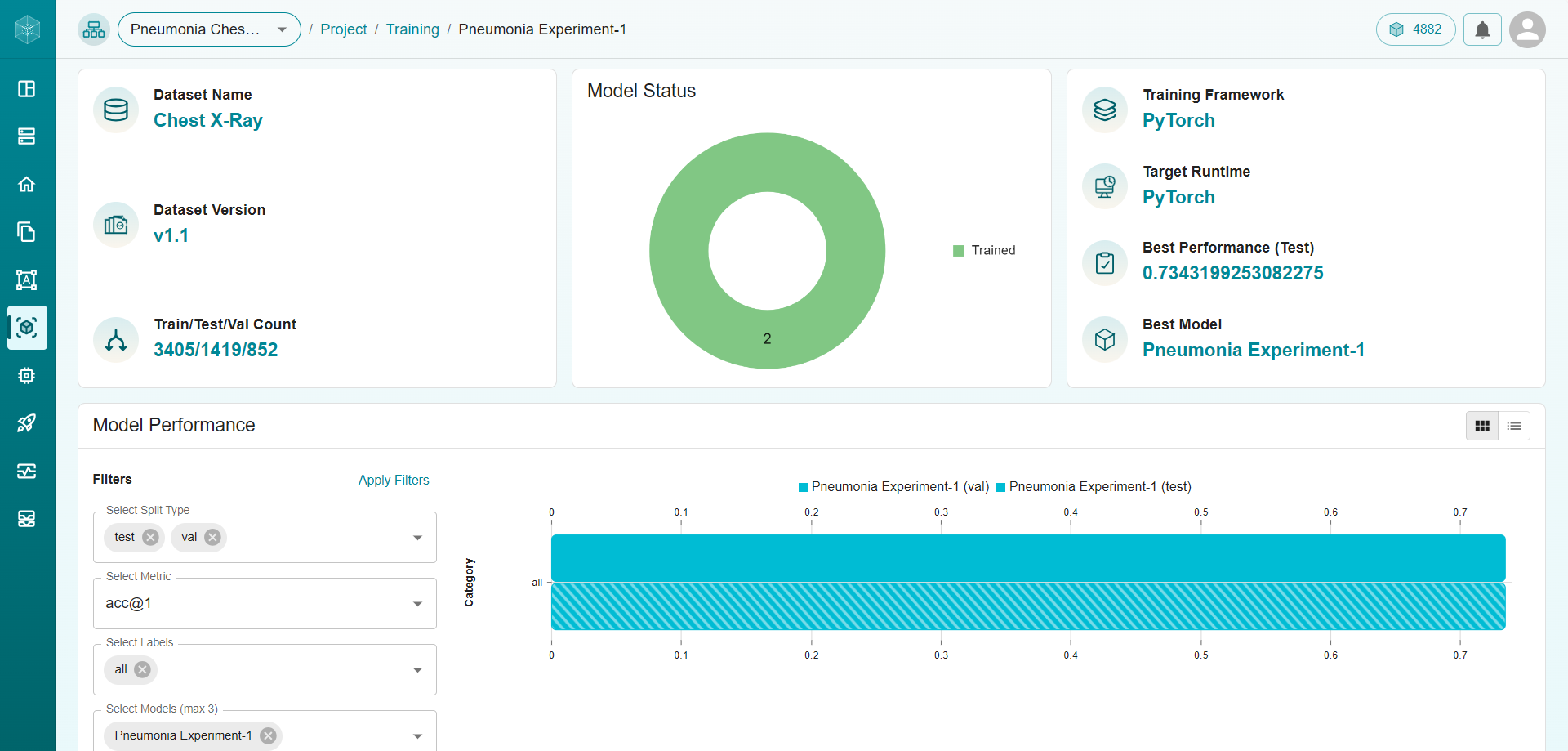
The DenseNet model was chosen for this application because of its ability to handle complicated picture categorization problems with high accuracy. Key setup information were included:
Batch Size: Set to 4, so that the model can handle smaller batches of chest X-ray pictures, which is better for memory management.
Epochs: Set to 80, which provides enough training cycles for the model to properly learn from the dataset.
Learning Rate: Set to 0.001 to optimize model learning speed while ensuring stable convergence.
Primary Metric: The performance was assessed using acc@5, which assesses the accuracy of the top five predictions and provides a more complete picture of the model’s performance.
Target Runtime: PyTorch was selected as the framework for training, providing for flexibility and efficiency. Matrice’s AutoML function improved model performance by tweaking hyperparameters, yielding a more accurate pneumonia classification model.
Step 4: Model Evaluation
Following training, the model’s performance was evaluated using a variety of metrics to ensure accurate pneumonia classification:
Accuracy: Evaluates the model’s overall ability to correctly categorize X-rays as either Pneumonia or Normal, considering both positive and negative cases.
Specificity: Indicates the proportion of true negatives (correctly identified normal X-rays) among all actual normal cases, helping to minimize false positives.
Precision: Measures the proportion of X-rays classified as pneumonia that are actually pneumonia, minimizing false positives and ensuring that positive classifications are reliable.
Metric |
Value |
|---|---|
Accuracy |
0.734 |
Specificity |
0.734 |
Precision |
0.500 |
These metrics confirm the model’s reliability in detecting and classifying pneumonia cases across diverse chest X-ray images.
Step 5: Model Inference
The model was ready for use in clinical settings after its efficacy was confirmed. The actions listed below were taken:
Export Formats: Supported formats such as ONNX and TensorRT were used to export the model, enabling integration with a range of medical devices and systems.
Compatibility for Real-Time Usage: These formats guarantee that the model can be integrated with clinical software and used on diagnostic devices, allowing for real-time pneumonia categorization and decision-making in medical settings.
The model may be readily integrated into current infrastructure by utilizing Matrice’s smooth export features, enabling quick deployment and enabling instant clinical impact.
Step 6: Model Deployment
Integrating the model with healthcare systems is simple because to Matrice’s safe and adaptable deployment options:
API Integration: Healthcare providers can easily integrate the pneumonia classification model into current workflows by using Matrice’s API to access it straight from radiography software or electronic health record (EHR) systems.
Scalable and Secure Access: Matrice’s cloud architecture guarantees that the model can grow to accommodate massive amounts of chest X-ray data while protecting patient privacy and data security in accordance with HIPAA and GDPR, among other healthcare norms.
Healthcare practitioners can use Matrice’s deployment capabilities to take advantage of the model’s predictions in real-time, which enhances clinical judgment and speeds up the time it takes for pneumonia patients to receive treatment.
Conclusion
Matrice’s AI-powered pneumonia classification model provides a scalable way to improve healthcare diagnostic speed and accuracy. Matrice gives healthcare providers a dependable tool to enable prompt, accurate diagnoses by automating the pneumonia detection process, which eventually improves patient care and treatment results.
Healthcare providers may use Matrice’s AI-powered solutions to optimize their diagnostic workflows, cutting down on time to diagnosis and guaranteeing that patients with pneumonia receive consistent, high-quality care.
Think CV, Think Matrice
Experience 40% faster deployment and slash development costs by 80%