Precision Anatomy: AI-Powered Liver Segmentation for Advanced Medical Imaging
Introduction
The liver is a vital organ, playing a central role in metabolism, detoxification, and numerous bodily functions. Consequently, it is susceptible to a wide range of diseases, including various forms of cancer (e.g., hepatocellular carcinoma, metastases), cirrhosis, and fatty liver disease. Accurate assessment of liver anatomy and pathology is paramount for effective diagnosis, precise surgical planning, targeted radiotherapy, and monitoring treatment response. Traditionally, these assessments rely on manual or semi-manual segmentation of medical images (like CT and MRI scans), a process that is highly time-consuming, prone to inter-observer variability, and often limits the quantitative analysis crucial for advanced clinical decisions.
Artificial Intelligence, particularly through cutting-edge computer vision and sophisticated image segmentation techniques, is revolutionizing medical imaging analysis. By enabling automated, precise, and consistent delineation of the liver and its internal structures from complex scan data, AI-powered liver segmentation promises to transform clinical workflows, enhance diagnostic accuracy, and optimize patient care pathways. This blog post explores our advanced AI model designed for robust liver segmentation, highlighting its technical capabilities, profound clinical benefits, diverse applications, and the significant impact it promises for advancing hepatology and surgical oncology.
1. The Critical Need for Automated Liver Segmentation
The urgency for efficient and accurate liver segmentation stems from several critical factors, directly impacting patient outcomes, clinical efficiency, and the precision of medical interventions:
Pre-surgical Planning for Liver Resection/Transplant: Accurate 3D models of the liver, including the identification of segments, tumors, and vascular structures, are essential for planning complex liver resections to ensure sufficient remnant liver volume or for donor-recipient matching in transplantation. Manual segmentation is arduous and can introduce errors.
Precise Radiotherapy Planning: For liver tumors, precise segmentation of the tumor and the healthy liver tissue is critical for accurate dose delivery, minimizing radiation exposure to healthy liver parenchyma while maximizing tumor eradication.
Quantitative Disease Assessment: Many liver diseases, such as fatty liver or cirrhosis, require volumetric analysis to assess disease progression or regression under treatment. Automated segmentation provides objective, reproducible volumetric data, far superior to subjective visual estimation.
Reduced Clinician Workload: Manual segmentation of liver volumes from hundreds of CT/MRI slices per patient is extremely labor-intensive and time-consuming for radiologists and oncologists. Automation frees up valuable expert time for more complex diagnostic and therapeutic tasks.
Improved Diagnostic Consistency: Automated segmentation reduces variability in measurements and classifications across different medical centers and radiologists, leading to more standardized and consistent diagnostic reports, which is vital for multi-center studies and clinical trials.
By addressing these multifaceted challenges, AI-powered liver segmentation is not merely a technological enhancement; it’s a fundamental step towards more precise, efficient, and ultimately safer patient management in hepatology and related medical fields.
2. Benefits of AI in Liver Segmentation
AI-powered liver segmentation systems offer a multitude of transformative benefits that are reshaping medical imaging and clinical decision-making:
Rapid and Accurate Volumetric Analysis: AI algorithms can delineate the liver contour across hundreds of CT or MRI slices in seconds, enabling fast and precise calculation of liver volume and tumor volumes. This speed is critical for time-sensitive pre-surgical planning.
Enhanced Precision for Surgical and Radiotherapy Planning: By providing pixel-level accurate 3D models of the liver, AI ensures that surgical resections are precisely planned to remove diseased tissue while preserving healthy liver, and that radiation doses are delivered with pinpoint accuracy to tumors.
Objective Disease Progression Monitoring: AI offers consistent, quantifiable metrics of liver and lesion volumes over time, enabling objective monitoring of disease progression, treatment response, and detection of subtle changes that might indicate recurrence or new lesions.
Reduced Inter-Observer Variability: Automated segmentation eliminates the inconsistencies inherent in manual tracing by different clinicians, leading to more standardized and reproducible measurements across studies and institutions.
Optimizing Clinical Workflow: Automating the arduous task of manual liver segmentation frees up radiologists, oncologists, and surgeons from tedious image processing, allowing them to focus on diagnosis, patient consultation, and therapeutic strategies.
Facilitating Research and Drug Development: By providing scalable and consistent quantitative imaging biomarkers, AI accelerates medical research into liver diseases, aids in the development of new therapies, and strengthens the statistical power of clinical trials.
3. Data Preparation for Robust AI
The success of our liver segmentation model is directly attributable to the meticulous preparation of a diverse and high-quality dataset. This process involved collecting and annotating vast quantities of medical images (primarily CT and MRI scans) featuring the liver, encompassing a wide range of patient anatomies and pathological conditions. Key aspects of our data preparation strategy included:
Multi-Modality Imaging: The dataset included scans from various imaging modalities (e.g., contrast-enhanced CT, non-contrast CT, MRI sequences like T1/T2-weighted) to ensure the model’s robustness to different image characteristics.
Diverse Patient Anatomy and Pathology: Images featured livers with varying sizes, shapes, and positions, as well as those affected by common pathologies such as tumors (primary and metastatic), cirrhosis, steatosis, and cysts, to train the model to segment healthy and diseased livers.
Varying Contrast Levels and Image Noise: The dataset incorporated scans with different contrast agents, acquisition protocols, and realistic levels of image noise or artifacts, mimicking the variability encountered in real-world clinical data.
Expert Pixel-Level Annotation: Highly experienced radiologists and medical imaging specialists meticulously delineated the boundaries of the liver parenchyma and, in some cases, specific lesions or vascular structures at the pixel level on each slice. This precise and detailed labeling served as the indispensable ground truth for supervised learning.
Rigorous Quality Control and Augmentation: Annotations underwent strict quality checks, and data augmentation techniques (e.g., intensity variations, noise injection, rotations, scaling) were applied to artificially expand the dataset’s diversity and enhance the model’s robustness.
Model Architecture
The foundation of our advanced liver segmentation system is the YOLOv9m architecture. While YOLO (You Only Look Once) is primarily known for its efficiency in object detection, its capabilities have been extended to perform instance segmentation. This allows for pixel-level delineation of objects, making it an ideal choice for the intricate task of precisely outlining internal organs like the liver within medical image slices. The YOLOv9m variant strikes an optimal balance between high segmentation accuracy and impressive inference speed, which is crucial for real-time clinical assessment and integration into imaging workflows.
Key advantages of YOLOv9m in the context of liver segmentation include:
Real-Time Segmentation of Medical Slices: YOLOv9m’s highly optimized design allows for extremely fast analysis of individual CT or MRI slices, enabling rapid generation of liver masks across an entire 3D volume.
Precise Boundary Delineation: The model excels at accurately segmenting the liver’s irregular and often subtle boundaries, distinguishing it from adjacent organs (e.g., kidney, stomach, spleen) and surrounding tissues at a pixel level.
Robustness to Anatomical Variability: Its sophisticated deep learning layers are highly effective at extracting intricate features from abdominal scans, ensuring reliable performance across diverse patient anatomies and disease states.
Efficient Mask Generation for Volumetric Analysis: Beyond just identifying the liver, YOLOv9m generates detailed pixel-level masks for each identified liver region on each slice, providing the precise output necessary for accurate 3D volumetric reconstruction and analysis.
Training Parameters
The model underwent extensive training to optimize its performance across the diverse dataset. The key training parameters were carefully selected to ensure stability, rapid convergence, and robust generalization to new, unseen medical images:
Parameter |
Value |
Description |
|---|---|---|
Base Model |
YOLOv9m |
The foundational deep learning architecture employed for the task, known for its efficiency and accuracy in instance segmentation. |
Batch Size |
8 |
Number of samples processed before the model’s internal parameters are updated, balancing training stability and computational efficiency. |
Learning Rate |
0.0005 |
Controls the step size during the optimization process, a conservative rate chosen for stable convergence and fine-tuning. |
Epochs |
70 |
Number of complete passes through the entire training dataset, ensuring the model learns extensively from the data and generalizes well. |
Optimizer |
AdamW |
An adaptive learning rate optimization algorithm (Adam with decoupled weight decay) known for its efficiency and strong performance in deep learning tasks. |
Inference Time |
~0.45s |
The average time taken for the trained model to process a single medical image slice and generate a liver segmentation mask. |
Model Evaluation
Our rigorous training and validation processes have yielded a model with robust capabilities for liver segmentation. The evaluation metrics below demonstrate the model’s precision, recall, and overall segmentation accuracy, proving its reliability for critical applications in medical imaging and clinical decision-making.
Metric |
Overall Performance |
Liver Tissue |
Surrounding Organs/Background |
|---|---|---|---|
Precision |
0.82 |
0.83 |
0.80 |
Recall |
0.81 |
0.85 |
0.78 |
F1 Score |
0.82 |
0.84 |
0.79 |
mAP |
0.71 |
0.75 |
0.68 |
Inference Time |
~0.45s |
- |
- |
Precision (0.82 Overall): This indicates that when our model identifies pixels as belonging to the liver, it is correct 82% of the time, minimizing false positives and ensuring accurate anatomical delineation.
Recall (0.81 Overall): With a recall of 81%, the model successfully identifies most of the actual liver pixels in an image. This is crucial for comprehensive volumetric assessment and ensuring all parts of the organ are captured.
F1 Score (0.82 Overall): The F1 Score, a harmonic mean of precision and recall, provides a balanced measure of the model’s overall accuracy in segmentation, reflecting its robust performance in both identifying and correctly outlining the liver.
Mean Average Precision (mAP) (0.71 Overall): As a comprehensive metric for instance segmentation tasks, mAP of 0.71 signifies strong overall performance across both “Liver Tissue” and “Surrounding Organs/Background” classes, indicating reliable accuracy in generating precise masks for medical image analysis.
Inference Time (~0.45s): The sub-second inference time ensures that liver segmentation can be generated almost instantaneously per slice, enabling rapid 3D reconstruction and analysis of entire patient scans.
The per-category metrics further highlight the model’s optimized performance: “Liver Tissue” demonstrates slightly higher recall, prioritizing the complete capture of the organ, while “Surrounding Organs/Background” shows good precision in differentiating other anatomical structures.
Epoch vs. Precision during Training
To demonstrate the training dynamics and performance stability of our model, the following graph illustrates the progression of precision over epochs. This visualization highlights how the model refined its accuracy during the training process, converging towards its optimal performance.
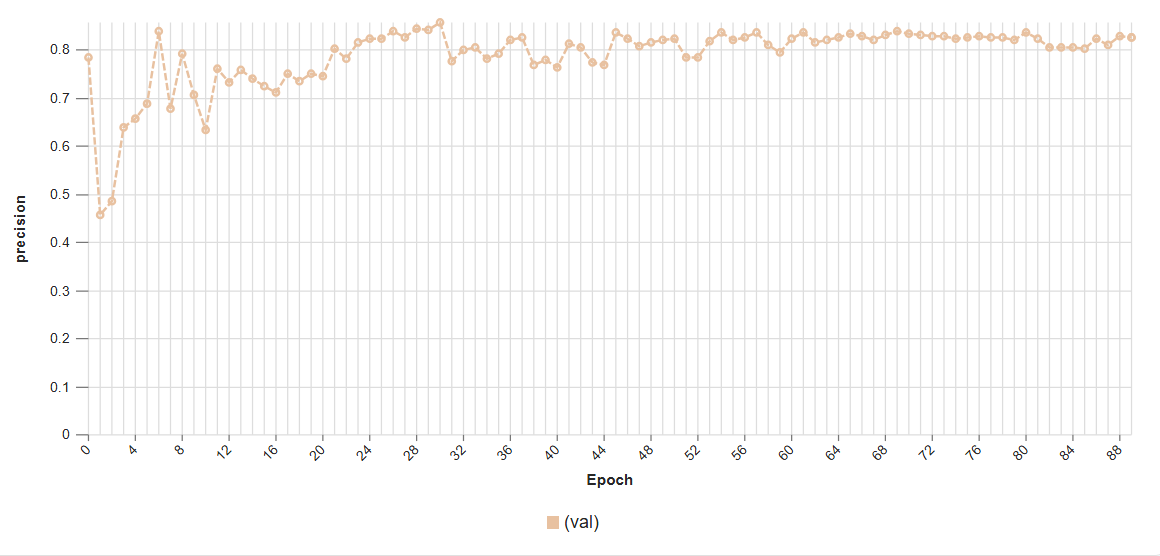 This graph illustrates the increase in model precision as training progresses over multiple epochs, showcasing the learning curve and stability of the YOLOv9m architecture in segmenting liver tissue.
Model Inference Examples
Below are conceptual examples demonstrating the model’s output when analyzing medical images for liver segmentation. The AI accurately identifies and outlines the liver organ, providing precise anatomical context for clinical use.
Example 1: Precise Liver Segmentation in a CT Scan Slice
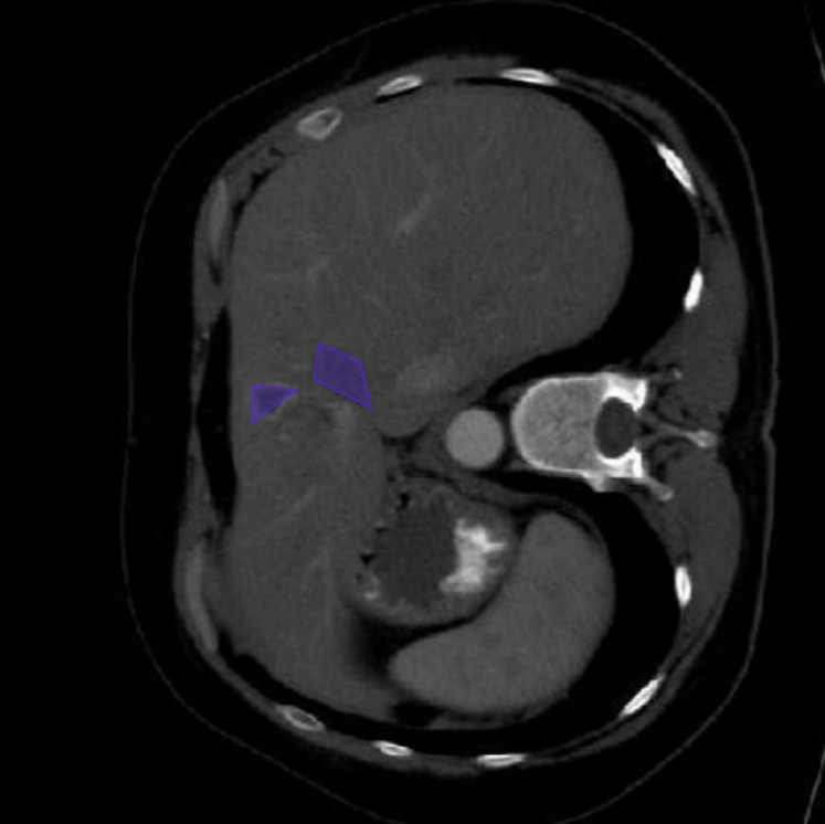 This image showcases our AI model in action, accurately delineating the liver (e.g., highlighted in green) within a complex abdominal CT scan slice. This precise segmentation provides the foundation for volumetric analysis and surgical planning.
Example 2: Liver Segmentation Across Different Imaging Modalities or Anatomies
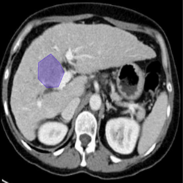 This example demonstrates the model’s versatility in segmenting the liver across varied patient anatomies or potentially different imaging modalities (e.g., MRI), highlighting its robust generalization capabilities.
4. Real-World Applications and Societal Impact
The deployment of this AI-powered liver segmentation system is poised to create a profound impact across various medical specialties and patient care pathways:
Pre-Surgical Planning and Navigation: Provides surgeons with accurate 3D volumetric models of the liver, its segments, and any tumors or vascular anomalies, enabling highly precise planning for resections, ablations, or transplant procedures.
Radiotherapy Treatment Planning: Facilitates the creation of highly accurate radiation plans for liver cancer patients, ensuring maximum dose delivery to tumors while sparing healthy liver tissue, thereby reducing side effects and improving outcomes.
Quantitative Assessment of Liver Diseases: Enables objective and reproducible measurement of liver volume, fat content (for fatty liver disease), or tumor burden, crucial for diagnosing, staging, and monitoring the progression of chronic liver diseases.
AI-Assisted Diagnosis and Reporting: Supports radiologists in rapidly reviewing and reporting on liver scans by providing initial automated segmentations, highlighting abnormalities, and quantifying relevant metrics.
Research and Clinical Trials: Provides a standardized and automated method for collecting high-throughput, quantitative liver imaging data for medical research, accelerating the development of new diagnostic biomarkers and therapies.
Liver Transplant Evaluation: Aids in the precise volumetric assessment of both donor and recipient livers, crucial for optimizing outcomes in liver transplantation.
5. Future Directions in AI-Powered Liver Segmentation
Our commitment to innovation ensures continuous development and enhancement of our AI capabilities in medical imaging. Future efforts will focus on:
Full 3D Volumetric Segmentation: Moving beyond slice-by-slice processing to develop truly 3D segmentation models that can process entire volumetric scans at once, potentially improving consistency across slices.
Sub-Segment and Lesion Segmentation: Granular segmentation of individual liver segments, and precise delineation of specific lesions (tumors, cysts) within the liver parenchyma, for more detailed diagnosis and intervention planning.
Multi-Organ Segmentation: Expanding the model’s capabilities to simultaneously segment multiple abdominal organs (e.g., kidneys, spleen, pancreas) within a single scan for comprehensive anatomical mapping.
Integration with Clinical Decision Support Systems: Embedding segmentation outputs directly into AI-powered tools that assist clinicians with diagnosis, prognosis prediction, and personalized treatment recommendations.
Real-time Intraoperative Guidance: Developing models capable of real-time segmentation and tracking of the liver during surgery (e.g., laparoscopic or robotic procedures) to provide augmented reality guidance for surgeons.
Conclusion
AI-powered liver segmentation represents a transformative leap forward in medical imaging and patient care. By delivering unparalleled accuracy, efficiency, and quantitative insights into liver anatomy and pathology, our solution empowers clinicians to make more precise diagnoses, plan more effective interventions, and ultimately improve outcomes for patients suffering from a wide range of liver conditions. As we continue to refine and expand these capabilities, the future promises an even more intelligent, data-driven, and personalized approach to hepatological care.