Cardiomegaly Detection in Chest X-rays: Enhancing Cardiac Diagnostics with Computer Vision
May 26, 2025
“From subtle radiographic clues to AI-assisted cardiac assessment—deep learning is transforming diagnostic imaging.”
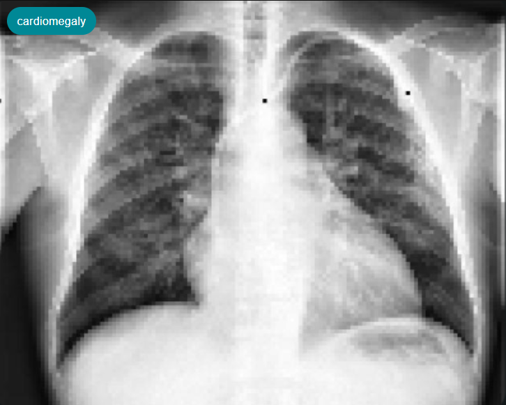
Cardiomegaly, an enlargement of the heart, is often a critical indicator of underlying cardiac conditions such as heart failure, hypertension, or valvular heart disease. Its timely and accurate diagnosis from chest X-rays (CXRs) is essential for guiding further investigations and appropriate patient management. While radiologists routinely evaluate CXRs for signs of Cardiomegaly, the assessment can be challenging due to subtle visual cues and workload pressures in busy clinical settings. Leveraging deep-learning-powered image classification, it’s now possible to automatically flag scans suggestive of Cardiomegaly within seconds, ensuring these cases receive prompt attention from clinicians.
This article walks through an end-to-end pipeline for developing such a system—from dataset preparation, model training and evaluation, to deployment strategies—highlighting how platforms like Matrice can streamline the creation of robust medical imaging AI solutions for cardiac assessment.
1. Why Accurate Cardiomegaly Detection Matters
The timely and accurate detection of Cardiomegaly is crucial for proactive patient care and efficient healthcare delivery:
Early Indication of Cardiac Issues: Identifying an enlarged heart can be one of the first signs of various cardiovascular diseases. Early detection allows for prompt further diagnostic tests (like echocardiograms) and the initiation of treatment, potentially slowing disease progression and improving long-term outcomes.
Supporting Radiologist Workload: In high-volume settings, radiologists analyze numerous images daily. AI pre-screening for Cardiomegaly can act as a vigilant assistant, highlighting potentially abnormal cases, thereby reducing fatigue, minimizing the risk of oversight, and improving overall diagnostic consistency.
Enhancing Care in Resource-Limited Settings: Access to specialist cardiological or radiological expertise may be limited in certain geographical areas. Automated AI tools for Cardiomegaly detection can bring a higher level of screening sensitivity to these locations, supporting local healthcare providers and facilitating better patient care, potentially through telemedicine.
Improving Diagnostic Standardization: AI models apply consistent criteria for assessment, unaffected by subjective factors, leading to more standardized preliminary findings related to heart size across different clinical environments.
2. Advantages of AI in Chest X-ray Analysis for Cardiomegaly
Integrating AI into the workflow for analyzing chest X-rays for conditions like Cardiomegaly offers significant benefits:
Benefit |
Impact |
|---|---|
Speed |
AI models can perform inference in under a second, enabling Picture Archiving and Communication Systems (PACS) to quickly flag studies suggestive of Cardiomegaly for prioritized review. |
Consistency |
AI algorithms deliver uniform assessment of cardiac size indicators across different shifts, sites, and patient populations, reducing inter-reader variability. |
Scalability |
Whether deployed in the cloud or on edge devices, AI solutions can scale to handle thousands of radiological studies daily, adapting to fluctuating demands. |
Cost-Efficiency |
By facilitating earlier identification of potential cardiac issues signaled by Cardiomegaly, AI can contribute to more timely management, potentially reducing long-term healthcare costs associated with advanced heart disease. |
Accessibility |
AI tools can democratize access to high-quality preliminary cardiac size assessments from CXRs, especially valuable in areas lacking specialized radiological expertise. |
3. Implementing Cardiomegaly Detection with MobileNetV2 (on the Matrice Platform)
Developing an effective AI solution for Cardiomegaly detection involves several key stages, from data handling to model deployment. This process can be significantly accelerated using comprehensive computer vision platforms like Matrice, which offer no-code or low-code environments for building and managing AI pipelines.
Dataset Preparation
A high-quality, well-annotated dataset is fundamental for training a reliable medical imaging AI model.
Name: Cardiomegaly Detection in Chest X-rays
Version:
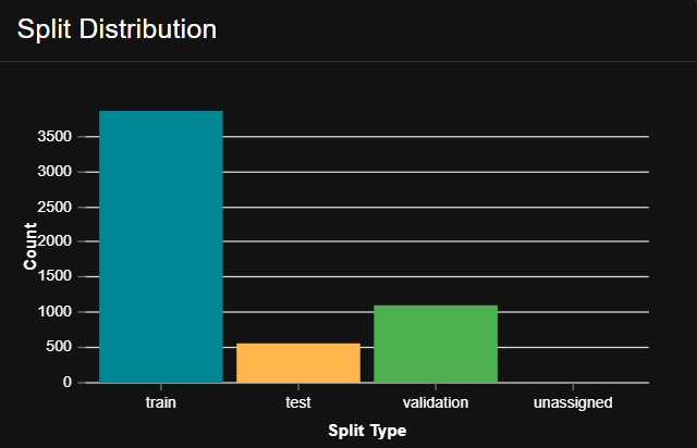
v1.0Total Images: 5,519 chest X-ray images.
Split:
Training images: 3,862
Validation images: 1,105
Test images: 552
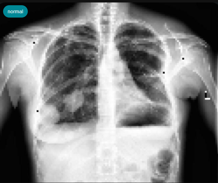
Classes: 2 classes for classification – Cardiomegaly (presence of an enlarged heart) and Normal (heart size within normal limits).
Format: Images are in standard PNG/JPG formats, processed as single-channel greyscale images, typical for CXRs.
Preprocessing: Standard medical image preprocessing steps, such as normalization and appropriate resizing, were applied to prepare the data for model training.
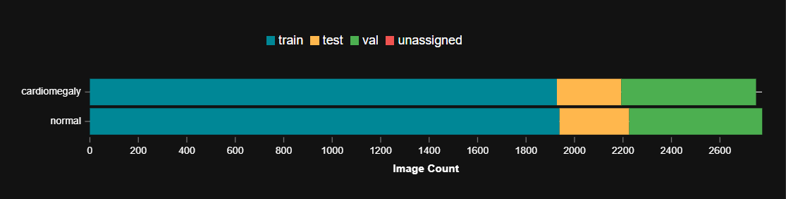 Number of samples within normal and cardiomegaly classes
Model Architecture: MobileNetV2
After benchmarking several architectures (including DenseNet-121 and ResNet-50), MobileNetV2 was selected as the primary model. This choice was driven by its excellent balance between diagnostic accuracy (particularly precision) and model size/computational efficiency for the Cardiomegaly detection task.
Model: MobileNetV2
Parameters: Approximately 3.5 Million, making it lightweight.
Key Strengths:
Designed for mobile and edge devices, offering low latency.
Utilizes inverted residuals and linear bottlenecks for efficiency.
Provides a strong performance baseline for various vision tasks, including medical image classification.
Training Framework: The model was trained using a PyTorch backend, often integrated within platforms like Matrice.
Training Parameters
The MobileNetV2 model was trained on the “Cardiomegaly Detection in Chest X-rays v1.0” dataset with the following configuration:
Hyper-parameter |
Value |
Notes |
|---|---|---|
Epochs |
70 |
Sufficient training iterations for convergence. |
Batch size |
8 |
Optimized for available GPU memory. |
Optimizer |
AdamW |
Momentum 0.95, Weight decay 0.0001 |
Learning Rate (LR) Schedule |
StepLR |
Initial LR 0.001, decaying to 0.0001 every 10 epochs. |
Loss Function |
Weighted Binary Cross-Entropy (BCE) + Focal Loss (γ=2.0) |
Addresses class imbalance and focuses on hard examples. |
Primary Metric |
Precision |
Prioritizes minimizing false positive detections. |
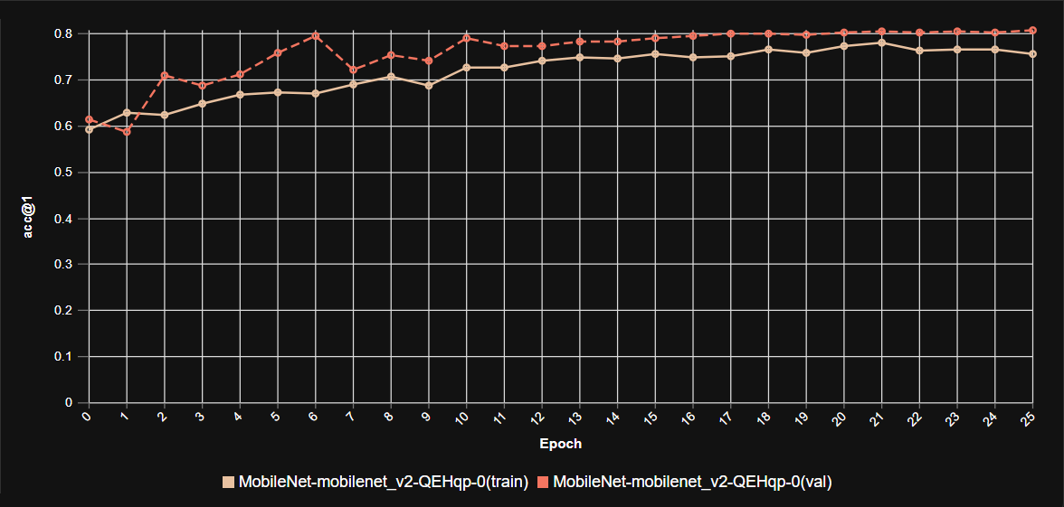 Training analysis
Model Evaluation
The performance of MobileNetV2 in detecting Cardiomegaly was compared against other architectures on the test set:
Model |
Parameters (M) |
Test Precision |
Test Recall |
Notes |
|---|---|---|---|---|
MobileNetV2 |
3.5 |
0.79 |
0.79 |
Selected Model |
DenseNet-121 |
8.0 |
0.59 |
0.64 |
Higher params, lower precision. |
ResNet-50 |
23.9 |
0.50 |
0.51 |
Much larger, lower performance. |
MobileNetV2 demonstrated high precision (0.79) and recall (0.79) for Cardiomegaly detection with a significantly smaller footprint (3.5M parameters), confirming its suitability for efficient and accurate screening, especially where deployment on edge devices is a consideration.
Deployment Strategy
The AI model for Cardiomegaly detection can be integrated into clinical workflows through various strategies:
API Microservice: Deploying the model as a containerized FastAPI (or similar) microservice that exposes a
/predictendpoint. This service can receive an X-ray image and return a classification label (Cardiomegaly/Normal) along with a probability score.PACS Integration: Developing a DICOM listener service that automatically receives new CXR studies from the PACS. The AI model analyzes these images, and findings suggestive of Cardiomegaly can be flagged and prioritized in the radiologist’s worklist for further review.
Edge Deployment: For rapid, on-site screening, the TensorRT-optimized model can run on portable X-ray carts or existing diagnostic workstations equipped with edge computing devices directly in clinics or hospital departments.
4. Real-World Impact and Clinical Benefits
The deployment of an AI-assisted Cardiomegaly detection system yields significant clinical and operational improvements:
Reduced Time to Flag Potential Issues: Integrating such an AI tool can cut down the initial screening time for potential Cardiomegaly from minutes to under 60 seconds per image, enabling faster routing for expert review.
Improved Detection Rates: AI assistance can help reduce the rate of missed findings for Cardiomegaly by a significant margin (e.g., data suggests up to 27% improvement in some scenarios for similar tasks), especially for subtle enlargements or in high-workload situations.
Enhanced Workflow Efficiency: Automating the initial screening allows radiologists and cardiologists to focus their expertise on confirmed or complex cases, optimizing their diagnostic workflow.
Feasibility for Resource-Constrained Hardware: The compact size of MobileNetV2 (model fits on < 50 MB of flash storage) makes it perfect for deployment on low-power, portable medical devices or existing hospital IT infrastructure.
Conclusion
AI-assisted Cardiomegaly detection using efficient deep learning models like MobileNetV2 demonstrates a powerful synergy between diagnostic need and computational capability. By leveraging structured datasets, optimized model architectures (often facilitated by platforms like Matrice), and robust deployment strategies, it’s possible to deliver actionable insights directly into existing clinical workflows. This technology not only saves critical time and resources but also enhances diagnostic accuracy and consistency, ultimately contributing to better patient outcomes in cardiac care.
Think CV, Think Matrice
Experience 40% faster deployment and slash development costs by 80%